Friday 23 September 2016

Some of the Conditions Knee Arthroscopy is Used to Treat and Diagnose

Arthrscopy of the knee is a common operation and there are more than 100,0000 Knee Arthroscopies performed every year in Australia. The procedure is generally a Day-Day hospital visit, performed often, though not always, under a general anaesthetic.


- Torn cartilage or meniscus: treated by trimming back the torn cartilage
- Loose, floating bodies: removal of loose cartilage or bone
- Knee-cap, (patello-femoral), disorders
- Washing infected knee joint
- General diagnostic purposes

Knee arthroscopy is most commonly recommended after you begin to consistently avoid regular lifestyle activities because of knee pain. Though, there are some medical conditions which can interfere with your doctor's decision to go ahead with arthroscopy.

Thursday 22 September 2016

Revolutionary Treatment For Total Hip Replacement Surgery

The Improvements made, since ivory was used in 1891 to replace the femoral heads of patients, have been numerous. Skin and other tissues, glass and then stainless steel, have all been used to replace degraded joint surfaces, in an attempt to make the movement of the hip joint smooth and pain-free.


In the 1960's Sir John Charnley, designed what he called a 'low friction arthroplasty', which was in principle the same as the prostheses used today. His prosthesis came in three parts; a metal femoral stem, a polyethylene acetabular cup, fixed with acrylic bone cement. The feature of 'low friction', Charnley managed to achieve by the smaller surface area of his femoral head design.

One of the great surgical advances over the last century is the total hip replacement. Total hip replacement revolutionised the treatment of hip ailments and is today one of the most successful, safe and reliable orthopaedic interventions in practice. 

Wednesday 27 July 2016

Arthroscopy Knee


Patients with certain types of meniscal tears and cartilage damage are prime candidates for the minimally invasive surgical intervention called Arthroscopy Knee diseased by osteoarthritis, rheumatoid or psoriatic arthritis can also be treated using arthroscopy.


The following covers the different types of treatments for meniscal tears and cartilage damage with a partial focus on arthroscopy assisted procedures.

Meniscal Tears

The most common non-surgical treatments of meniscal tears are:
  • Rest, Ice, Compression, Elevation, (RICE)
  • Non-Steroidal anti-inflammatory medicines
The outer third of the meniscus has a healthy supply of blood so can heal successfully without the trimming of smaller tears using arthroscopy. Though the inner two thirds of meniscus plates have a much reduced blood supply meaning tears in the region are more likely to require arthroscopic intervention.

For surgical treatment of meniscal tears there is:
  • Arthroscopy: a miniature camera is inserted through a small incision to provide a view of the inside of the knee. Miniature surgical instruments are then inserted in separate incisions to trim or repair the tear.

Cartilage damage


As cartilage does not heal easily of its own accord, surgical techniques have been developed to assist cartilage regrowth. Cartilage restoration can relieve pain and delay the onset of arthritis. Arthroscopy has many different applications in treating cartilage damage, such as:

  • Microfracture: A small pointed tool called an awl is used to create small holes in the subchondral bone. This stimulates new blood supply and cell growth in the cartilage.
  • Drilling: stimulates the production of healthy cartilage. Multiple holes are made in the subchondral bone using drills or wires to initiate a healing response. Though the tools used are less precise than microfracture. Arthroscopy.
  • Abrasion Arthroplasty: instead of using drills or wires, high-speed burrs remove the damaged cartilage and reach the subchondral bone. Arthroscopy.
  • Autologous Chondrocyte Implantation: using arthroscopy of the knee this procedure extracts healthy cartilage tissue, then grows the cells in a laboratory over 4-5 weeks. Using open knee surgery, the cells are the re-implanted in the site of the damaged cartilage. Arthroscopy and arthrotomy, (open surgery).
  • Osteochondral Autograft Transplantation: healthy cartilage is lifted using a cylindrical plug from a non-weight-bearing area of the knee joint, then transferred to the defective area and pressed into place. A single plug or multiple plugs can be used. Arthroscopy and open surgery.
  • Osteochondral Allograft Transplantation: when the defect is larger, instead of autografts, allografts use tissue grafts taken from cadaver donors. Allografts usually use open knee surgery.

Monday 11 July 2016

Orthopaedic Surgeon brief on sub-specialties depending on the region of the body


Orthopaedics is a branch of medical surgery and therapy that treats injuries and diseases of the body’s skeletal system. This includes the bones, joints, muscles, ligaments and tendons. This vast system that enables the uprightness and movement of the human frame is called the musculoskeletal system.
When first practiced orthopaedics treated children with spinal, leg or arm deformities. Though orthopaedic surgeons now help patients of all ages and for a range of different conditions.
Within orthopaedics there are many different sub-specialties depending on the region of the body they treat. Orthopaedic surgeons can specialise in:
Hand surgery

Hand surgery treats a range of conditions from cuts, burns, crushing injuries or diseases of the hand. Some common conditions are congenital deformities; repetitive stress injuries, like carpal tunnel syndrome; arthritis; and tendon repair. The priority for orthopaedic surgeons is to adequately reconstruct the skin, bone, nerves, tendons and joints. This is to protect the anatomy of the hand for recovery of functionality and aid the healing process by limiting the chance of infection. Early intervention is very beneficial.

Shoulder and elbow

The shoulder is a complex and unstable joint which can get injured easily. The elbow is vulnerable to deterioration from age or injury related conditions as well. The orthopaedic surgeon has many means of treating the muscles, connective tissue, or damaged joints that come from the overuse or injury of the shoulder or elbow. Some common procedures are arthroscopy or arthroplasty.


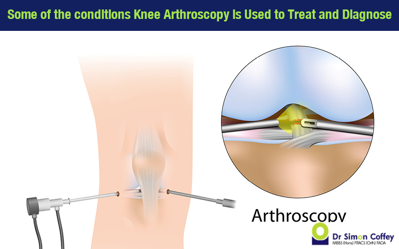
Arthroscopy is a form of keyhole surgery that magnifies and creates a visualisation of the interior of the body around the joint being operated on. While arthroplasty is the surgical removal and replacement of a joint or a section of the joint.

Hip and Knee

The hips and knees are the largest joints in the body. They are designed to work in close coordination to provide the mobility most people take for granted. The hip is a large ball and socket joint susceptible to both injury and disease. Some of the most common forms of hip disease are:

  • Osteoarthritis
  • Rheumatoid arthritis
  • Post-traumatic arthritis
  • Avascular necrosis

As with the shoulder and elbow, both the knee and the hip can be successfully treated by either arthroscopy or arthroplasty. Hip and knee replacement are two of the most common and successful procedures available to patients.
Pediatric orthopaedics
The pediatric orthopaedic surgeon specialises in children’s musculoskeletal problems. As children are still growing, the body's response to injuries, infections, and deformities can be quite different from the recovery of adults. Some of the conditions particular to this specialty are:
  • Limb and spine deformities, (clubfoot, scoliosis, limb length differences)
  • Gait abnormalities (limping)
  • Broken bones
  • Bone or joint infections and tumors
Foot and Ankle surgery

The treatment of the the foot and ankle is a sub-specialty of both orthopaedics and podiatry. The surgeon treats conditions that range from:
  • Trauma: such as fractures to the malleolar, tibial pilon, calcaneus, navicular, metatarsal, phalangeal
  • Arthritis: joints of the tarsals, metatarsals and phalanges, etc.
  • Congenital and acquired deformities: flatfoot, non-neuromuscular foot deformity, diabetic foot disorders, hallux valgus and several common pediatric foot and ankle conditions.
Spine Surgery

The are many potential causes for the need for spinal surgery. There is aging, improper body mechanics, trauma and structural abnormalities. These conditions can lead to back pain and symptoms such as leg pain and/or numbness or leg weakness. The most common procedures conducted are:

  • Discectomy or Micro-discectomy: removal of a herniated intervertebral disc.
  • Laminectomy: removal of the thin bony plate on the back of the vertebra called the laminae.
  • Laminotomy: removal of a portion of the vertebral arch, (lamina), that covers the spinal cord.
  • Foraminotomy: removal of bone or tissue at/in the passageway where nerve roots branch off the spinal cord.
  • Disc replacement: the injured disc is replaced with an artificial one.
  • Spinal fusion: technique used to join two vertebrae.


The three sub-specialties of orthopaedics not based on the body region where they occur are:
  • Musculoskeletal oncology, (cancer treatment)
  • Surgical sports medicine, (activity based injury)
  • Orthopaedic trauma, (accident based injury)

Wednesday 13 April 2016

Hip Replacement Recovery

In large part, the pace and quality of your recovery post a total hip replacement is down to how well you follow the advice given by your surgeon. The first weeks after an operation are crucial to the success of your recovery and the advice you are given should be followed.


Sometimes feeling well can put you in a state of mind where you are less likely to be cautious. This can lead to unnecessary accidents and setbacks, but equally, being too cautious can limit the return of your flexibility and your capacity for activity overall. Provided you are not taking risks the exercises should be done routinely even if you are feeling a little unwell.


There are many aspects to your homecare regimen. The first concern of hip replacement recovery is taking care of the healing process of the incision site. Usually, at around two weeks the stitches or staples will be removed. During this first fortnight, it is very important to keep the area dry at all times. Special waterproof patches can be bought, partners or relatives can help, or you can organise general homecare assistance.
Some people overlook that during their hip replacement recovery they might experience a loss of appetite. This reaction can last several weeks into the recovery process and if not countered can contribute to poorer recovery rates. The main reason is that a good diet promotes the healing process and will restore muscle strength. Often the loss of muscle strength is a contributing factor to any unwillingness to perform the necessary exercises crucial to regaining mobility. Iron supplements are also recommended.

The exercises you are assigned to do during your hip replacement recovery are particularly important. For the first few weeks post surgery, the care and regularity of your exercises will contribute to its overall success.

Normally, patients will have a customised exercise routine recommended by their doctor, but some of the more common exercises are:

  • A graduated walking program to slowly increase mobility
  • Slowly taking on normal household activities; like sitting, standing, and climbing stairs
  • Tailored exercises several times a day.
These exercises restore movement and strengthen your hip. A lot of people opt for the services of a physical therapist. They can assist you at home for the first few weeks after surgery, or there is also the option of choosing the services of a therapy center instead. In fact, the role of rehabilitation centers in a patients hip replacement recovery can be instrumental, in the rate of recovery and the patients proper understanding of the ‘do’s and don’ts’ of the recovery process.

The standard resumption of light daily duties usually takes anywhere in between 3-6 weeks, with the possibility of discomfort during activity and at night.

Total Hip Replacement

Total Hip Replacement is an operation to remove the acetabulum and femoral head of the hip joint to implant a ball and socket prosthesis to function as a moveable joint as a replacement. Total Hip Replacement is called Arthroplasty.


In a well-functioning hip joint both the acetabulum and the femoral head are lined with surface cartilage, or what is called articular cartilage. This surface layer of cartilage is encapsulated by synovial fluid which acts as a lubricant to assist joint movement. The cartilage acts as a shock absorber and smoothens the roll of the joint and under normal circumstances lasts a lifetime.


When the depletion of cartilage exposes bone to friction, chemical reactions and inflammation occur to degenerate the condition of the cartilage further. The mechanical wear caused by bone on bone friction is the primary condition of osteoarthritis and the source of the associated swelling and pain. 

The prosthesis used in Total Hip Replacement comes in two sections. The upper thighbone, (femoral head), implant is generally made of a metal or ceramic ball attached to a splint inserted into the top of the bone. The socket half of the implant is made of ceramic or a special plastic called polyethylene. Due to the acetabulum being concaved the implant is surrounded by bone surface to adhere to.

As to how the implants are affixed, two standard types of total replacement methods exist. There is the cemented method and the cement-less. The cement used to affix implants to bone is called methyl methacrylate. The cement-less method however uses microscopically porous materials to allow the bone to grow into and adhere to the implant.